Sudden hearing loss can be frightening and confusing, often leaving patients and doctors searching for answers. Researchers at the Keck School of Medicine of USC have adapted a common eye imaging tool, Optical Coherence Tomography (OCT), to see into the inner ear, offering a promising new method for diagnosing and treating hearing problems quickly and accurately.
Why it matters
Adapting OCT for the inner ear could accelerate the diagnosis of inner ear fluid imbalances, a key factor in conditions like Ménière’s disease, resulting in faster, more targeted treatment for sudden hearing loss and vertigo.
“Patient symptoms—sensations of hearing loss, pressure in the ear, or vertigo—can be affected by many unrelated factors such as mood, stress, nasal congestion and allergies. However, inner ear fluid levels reliably assess what is going on inside the inner ear. We have lots of treatment options available, but it often takes many weeks to figure out what works for a given patient. This could be a faster way to help.” —Lead author John Oghalai, MD, professor and chair in the Caruso Department of Otolaryngology – Head and Neck Surgery and the Leon J. Tiber and David S. Alpert Chair in Medicine at the Keck School of Medicine.
What is endolymph?
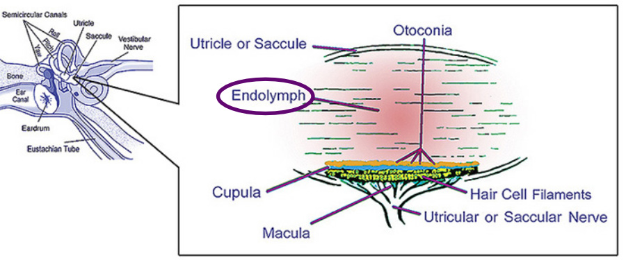
Endolymph is a sensory fluid in your inner ear that plays a role in both your hearing and balance (vestibular) systems. Its movement triggers nerve cells within your inner ear to communicate information about sounds and body position to your brain. Too much endolymph in your inner ear may lead to Ménière’s disease. —Cleveland Clinic
Where is endolymph in the inner ear?
Endolymph fills a network of ducts in the inner, the membranous labyrinth, which includes the cochlear duct (where sound waves are converted into auditory signals) and the organs that sense movement—the utricle, saccule and semicircular canals.
The problem
Conditions like Ménière’s disease involve a buildup of fluid (endolymph) in the inner ear, causing hearing loss and dizziness. Current diagnostic tools like MRI lack the detail needed to reliably visualize these fluid levels, leading to delays in treatment.
How it works
OCT, which uses light waves to create detailed 3D images of tissue (similar to how ultrasound uses sound), was used during ear surgeries on 19 patients. After temporarily removing a bone, researchers successfully imaged the inner ear's fluid compartments.
By the numbers
- The study included 6 patients with normal hearing, 4 with Ménière’s disease, and 9 with a benign tumor (vestibular schwannoma).
- OCT scans revealed that patients with Ménière’s or a tumor had significantly higher endolymph levels
- The higher levels of endolymph correlated directly with the degree of their hearing loss.
What to know
Measuring endolymph accurately has been a major challenge—OCT provides that crucial measurement.
“We’ve known for a long time that endolymph is related to hearing loss, but until now, measuring it in a living patient has been a major challenge.” —Dr. John Oghalai
The bottom line
Compared to MRI, OCT is faster, less expensive, and could allow immediate testing of treatments. For example, imagine an image taken, a medication given, and another image taken 30 minutes later to see if it worked.
The takeaway
This innovation repurposes OCT to address a significant gap in hearing care, promising quicker answers and better results for patients dealing with sudden and unexplained hearing loss.
Schedule a free hearing screening
Learn about the health of your hearing with a free 15-minute hearing screening by an audiologist.
★ Call 708-599-9500 to schedule your free screening.
★ For facts about hearing loss and hearing aid options, grab your copy of The Hearing Loss Guide.
★ Sign up for our newsletter for the latest on Hearing aids, dementia triggered by hearing loss, pediatric speech and hearing, speech-language therapies, Parkinson's Voice therapies, and occupational-hearing conservation. We publish our newsletter eight times a year.
Don't let untreated hearing loss spoil your enjoyment of life.
Crest Hill, IL - 630-633-5060 | Palos Hills, IL - 708-599-9500